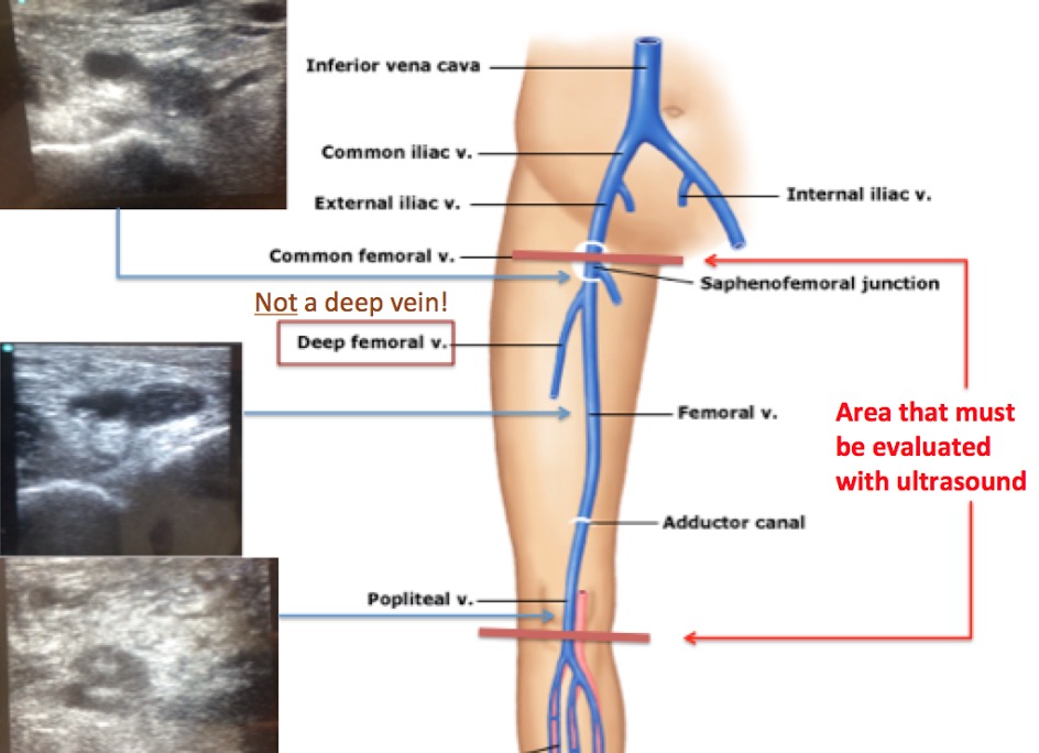
These ultrasounds performed by R1 Emergency Medicine Resident LM at HUM nicely demonstrates a very large DVT extending from the saphenofemoral junction to the popliteal vein. Remember that when looking for a DVT with bedside ultrasound, we must always evaluate the deep veins from where the saphenous vein enters the femoral vein near the inguinal area down to the popliteal vein. Dr. LM correctly did this and found that the deep venous system was clotted the entire way down.
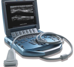
When performing a DVT study, start by placing the probe (linear probe is usually the best choice, however this can be done with an abdominal probe and if the patient is very large, the depth offered by the abdominal probe may be required) on the proximal thigh and find the femoral vein.
Scan up and down until you find the saphenous vein entering the common femoral vein medially. This will be your starting point. Although the saphenous vein is a superficial vein, if there is a clot in the saphenous vein, it can extend into the femoral vein and so must be treated the same as a true DVT.

Once the saphenous vein is located, put pressure on the probe in an attempt to fully collapse the veins. Exert as much pressure as you need to be confident that the vein is either fully collapsing (normal) or will not collapse (thrombus present). In our patient, there is a large clot in the common femoral vein, as it does not collapse at all. In the picture above, the clot is quite obvious even before compression is applied however in some patients, you will not be able to see the clot, but will only discover the presence of one when the vein fails to collapse. You will see in the video that the saphenous vein does collapse.
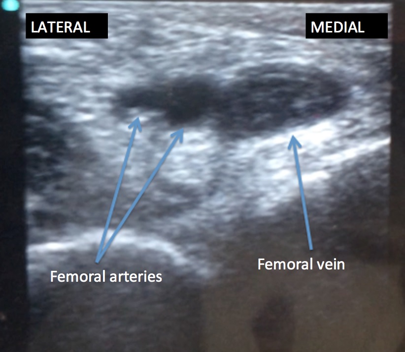
Continue the study by slowly scanning down the patient’s thigh, following the femoral vein distally. Every inch of so, put pressure on the probe in order to ensure that the femoral vein will collapse. As you scan, you will see the deep femoral arteries running laterally to the femoral vein.
You may also see the deep femoral vein, or profunda femoris, converge with the femoral vein. Interestingly, despite it’s name, the deep femoral vein is not thought to be a source of emboli and therefore is not part of the deep venous system that we must evaluate. On the contrary, some sources call the femoral vein that we must evaluate the “superficial” femoral vein, yet this superficial vein is part of the deep system. So the superficial vein is actually deep and the deep vein is actually superficial! Why doctors created such confusing names, I have no idea, but no worries, just follow the largest vein as it moves down the leg, again, pressing hard with the probe every inch or so to make sure the venous system fully collapses.
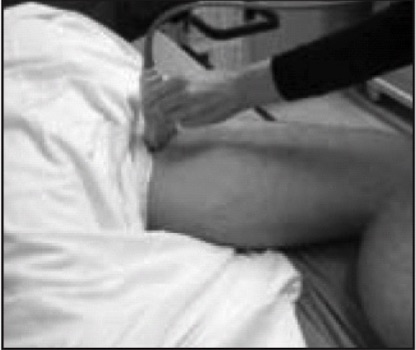
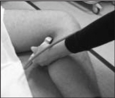
Once you have scanned through the distal thigh, the last thing you must do is look at the popliteal vein. Do this by placing the probe behind the patient’s knee. You may need to support the patient’s leg with the hand you are not scanning with.
From this position, you should see the popliteal vein on top of the popliteal artery.
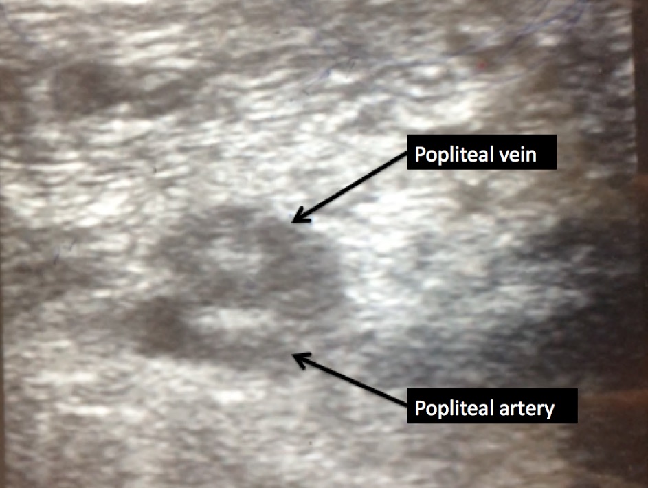
Again, press hard in an attempt to fully collapse the popliteal vein. Once you have done this, you have finished the study, and hopefully for the patient ruled out a DVT. Our patient was not so lucky, when you watch the videos, you will see that the venous system does not collapse at any point.
The patient was diagnosed with a large DVT, placed on enoxaparin and admitted to internal medicine for further management.
For reference, here is a link to a video which shows a normally compressible femoral vein, or a vein in which there is no DVT.
And here is the link to the website where the video comes from, which also has other very good examples of both positive and negative DVT studies.
Great work.