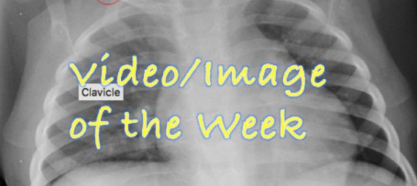
A 2 yo male was brought to the HUM ED for fever and facial swelling that began 4 days prior after a fall while playing. On exam, the patient had swelling of the scalp and right frontal region and was unable to open his right eye.
The EM docs obtained the following x-ray during the workup and were concerned for child abuse due to findings on the bilateral collar bone:
Are these findings the result of past trauma??
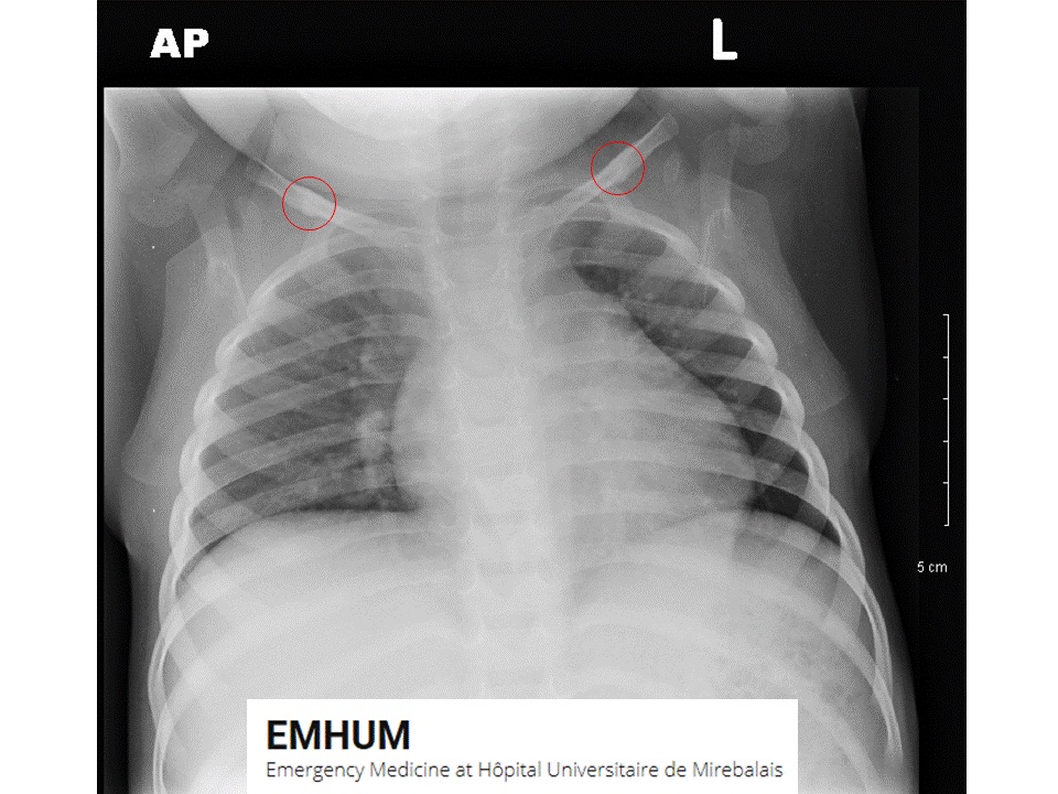
No! This was not a case of child abuse. While multiple unexplained fractures in young children should raise suspicion of child abuse, these findings are the result of normal ossification of the clavicles, and in this case, trauma was a distraction from the real disease process that needed to be addressed: a scalp abscess.