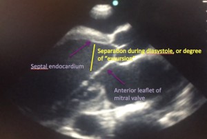
This is a parasternal long view of the heart performed on a cardiac patient in the internal medicine ward at HSN by a 1st year family medicine resident. It shows significantly depressed left ventricular function with very poor ejection fraction.
In a NORMAL heart, in the parasternal long view, the anterior leaflet of the mitral valve (see labeled picture below) can be seen touching or almost touching the septal endocardium in early diastole. The amount of separation, or “degree of excursion,” correlates to the contractility of the left ventricle. The larger the separation, the worse the contractility. Notice how in this heart, the separation is very large, consistent with very poor left ventricular dysfunction.
Notes from our cardiologist:
In case you have never seen a “normal” heart, here is an echo clip of a normal patient.
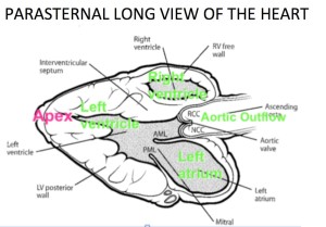
The key features to evaluate on the PARASTERNAL LONG AXIS view include:
1. Left ventricle systolic function: Normally, the walls of the heart move towards each other by about 25%. This is called the “fractional shortening”
2. Right ventricle size: the diameter of the right ventricle should only be up to 2x the dimension of the aorta
3. Mitral valve: leaflets should be thin with good mobility – ruling out severe mitral stenosis
4. Pericardium: normally, there is no pericardial effusion.


