ekg-signal-md-1

EKG – 1 – Complete Heart Block

64 year of woman presents to HUM after an episode of dizziness which caused her to fall.  No LOC consciousness, no traumatic injury from the fall.  Patient without symptoms in the ED.

Vitals:  SBP 140, HR 27, RR 14, O2 Sat 99% on room air.  Exam unremarkable, appears well for age.  Physical exam only demonstrates bradycardia.

What does her EKG show?
Screen Shot 2016-01-31 at 4.25.08 PM


This EKG shows a THIRD DEGREE, or COMPLETE HEART BLOCK, meaning there is no communication between the atria and the ventricules.

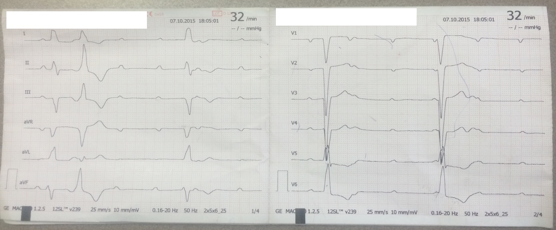
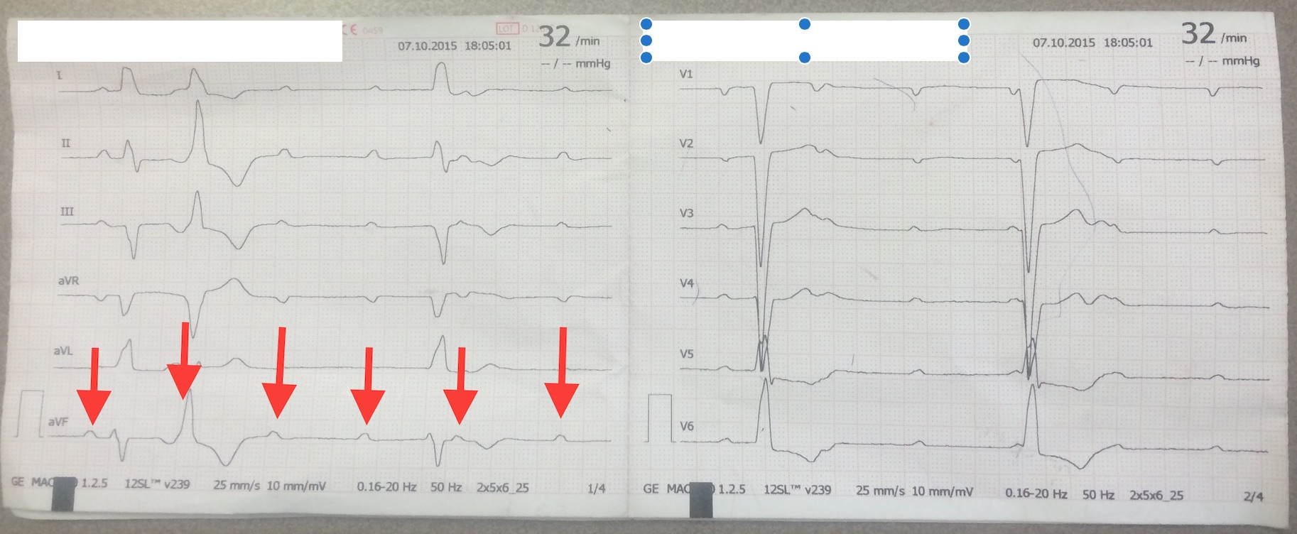
AXIS:  around – 30% – slight left axis deviation (I+, III -, II isoelectric)

RATE: around 30 bpm

RHYTHM:  Regular w/ p waves without QRS complex = heart block

INTERVAL:  QRS wide, there are two ventricular escape beats (remember every heart cell can be a pacemaker the ventricular is just slower, so if no electrical conduction comes from the atria the ventricular will have these escape beats) and one PVC.
Size:  unable to determine

ST SEGMENTS: unremarkable


Since there are p waves without QRS waves, this must be a second degree or third degree block.  Because the PR intervals ARE NOT always the same, this cannot be a mobitz type II block which has the same PR intervals when present.  Looking closely, you can see that the p waves are going independently without relation to the QRS (see second EKG which shows the p waves).
 Screen Shot 2016-01-31 at 4.25.50 PM
 This patient has a stable third degree heart blocking, best management would be observation on a monitor until pacemaker can be placed.  In our case, this was not an option at HUM so we referred her to a hospital in the south where she can get a pacemaker.


Share

Leave a Reply

Your email address will not be published. Required fields are marked *