
Dr. Menager, PGY-1 EM resident at HUM, performed this ultrasound on a young woman who presented with dysuria and right-sided constant flank pain. The patient’s vital signs were stable, pain was controlled in the ED and urinalysis was consistent with infection. Her overall presentation was highly suggestive of pyelonephritis and preparation was made for discharge with outpatient followup. Dr. Menager however cleverly performed an ultrasound prior to discharging her and found a large obstructive, infected renal stone. Instead of being discharged, the patient appropriately remained in the ED for IV antibiotics. A CT and urologic consultation were obtained. Her CT is below:
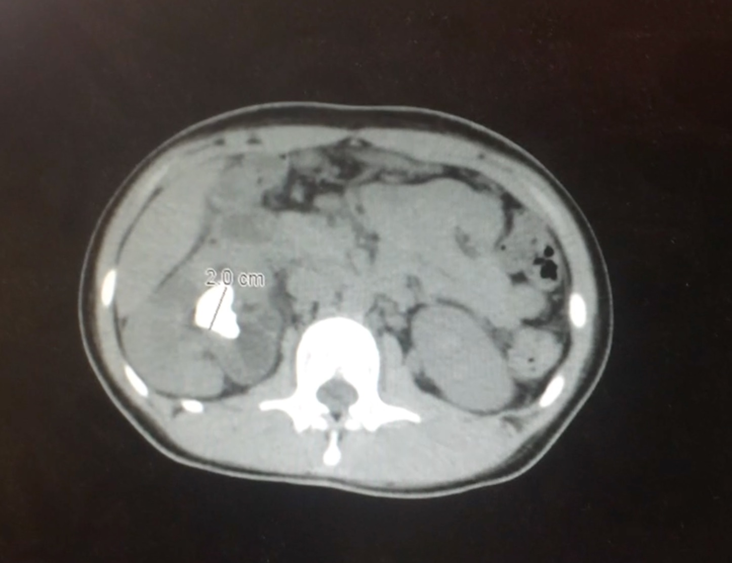
Large staghorn calculus in right kidney
This case highlights why it is so important to ultrasound every suspected pyelonephritis prior to discharge to ensure there is no concerning hydronephrosis. While stable pyelonephritis can be discharged with antibiotics and good followup, an infected and obstructed kidney stone is a urologic emergency!