R2 EM resident Dr. Duneant, accompanied by one of our visiting professors, Dr. Aimee Tang, International Emergency Health Fellow at North Shore-LIJ, performed this ultrasound on a 2 yr old boy who was referred to the HUM ED from an outside hospital.
The boy fell one week prior from a height of about 1 m. He had a short witnessed loss of consciousness followed by 2 episodes of brief tonic-clonic seizure-like episodes right after the fall. His parents brought him to a small community hospital where he was observed for one day without further seizure activity or changes in neurologic status. He was sent home and remained at baseline however one week later parents noticed a mass on the child’s head and went back to the hospital and was referred to HUM.
On examination, the patient was found to have an approximately 4×3 soft boggy mass overlying the left parietal bone. There was no evidence of abrasion or bleeding. Patient appeared well, interacting normally with parents and examiners.
The ultrasound performed by Dr. Duneant shows a significantly depressed skull fracture which was not able to be palpated through the overlying hematoma. Skull fractures, like all bone fractures, can be found on ultrasound by looking for a break in the cortex of the bone. 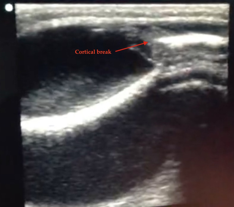 Bone, whether in the hand or the head, appears as a linear hyperdensity. To find a break, you must follow the cortex of the bone in either the longitudinal or transverse view until you see a break in the “white line” that is the bone.
A 2013 article in Pediatrics suggests that ultrasound can diagnose pediatric skull fractures with moderate sensitivity (88%) and high specificity (97%). Using ultrasound to diagnose bone fractures is a relatively new concept, however in the last five years, there is increasing awareness of the utility of ultrasound to help in the diagnosis and management of all sorts of fractures including clavicular, rib, sternal, long bone, and distal forearm fractures.
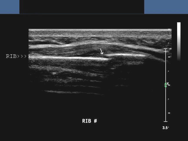
Example image of a rib fracture from ultrasoundconnection.com
More investigations will need to be undertaken to determine the true utility of bedside ultrasound in acute fracture management and we are sure to see studies addressing this topic coming out in the next few years. But certainly at this point, if your x-ray machine is not working and you don’t have access to a CT scanner, remember that fractures are just another thing that bedside ultrasound can help you “see” as you do your best to help your patient.
Here is the CT the HUM ED team obtained confirming the depressed skull fracture and showing a small subdural bleed adjacent to the depressed fragment:
The fracture was closed and so antibiotics were not warranted and were not given. The patient was started on anti-epileptics and referred to a neurosurgeon in Port-au-Prince.
More information on how to ultrasound fractures: