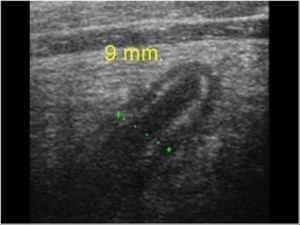
A 25 yr-old female presented to HUM with RLQ pain and nausea. Megan Mickley, one of our awesome visiting professors, Ultrasound Fellow from Children’s Hospital of Philadelphia, performed this ultrasound with one of our residents, which nicely demonstrates acute appendicitis.
How to perform an ultrasound to evaluate for appendicitis:
Take the LINEAR probe, place it over the abdominal wall at the area of maximal tenderness. Exert gentle pressure with either the ultrasound or your free hand. This is what’s called “graded compression.” This does two things. First, it pushes away bowel gas and decreases the distance between the appendix and the probe allowing you to visualize the structures better (make sure you give the patient adequate pain medication so they will tolerate this!). Second, it allows you to determine the compressibility, or in the case of an inflamed appendix, the NONCOMPRESSIBILITY of the structures you are looking at.
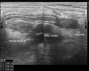
Case courtesy of Dr Maulik S Patel, Radiopaedia.org. From the case rID: 16327
Hopefully, if you place it over the area of maximal tenderness, which often is where an inflamed appendix most closely approximates the peritoneum, you will quickly be able to visualize the appendix. Often however, you will need to carefully search that area with your probe looking for a noncompressible, tubular structure with a blind end. The psoas muscles and illiac vessels can be used as landmarks, as the appendix usual lies anterior to these structures.
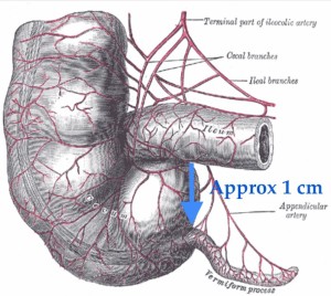
If you see a nonperistaltic structure containing gas and fluid that does not end, that is the ascending colon. If you see this, move the probe inferiorly and try to identify the terminal ileum, a compressible structure with peristalsis. You can then try to find the appendix, which usually arises approximately 1 cm inferior to the terminal ileum. Once you find the appendix, remember to measure it from OUTER wall to OUTER wall.
The diagnostic criteria for appendicitis on ultrasound are:
- Blind-ended, tubular, noncompressible, aperistaltic structure in the RLQ
- Diameter > 6 mm,
You may also be able to find:
- Appendiceal wall hyperemia as seen on color Doppler.
- Appendicoliths, which appears as a hyperechoic foci that casts an anechoic shadow
- Pericecal inflammatory changes such as free fluid or hyperechoic fat (fat stranding).
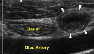
In cross section, a distended appendix will have a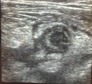 target like appearance, just as seen in the ultrasound performed by Dr. Mickley (pictured to the right). Remember you must try to visualize the entire appendix in a longitudinal plane in order to ensure that it is a blind ending structure, meaning that it ends. If you just look in a cross sectional plane, or do not visualize the end of the appendix in a longitudinal plane, it is very easy to incorrectly think that the terminal ileum is the appendix. From the picture below (distended appendix has white arrows), it is easy to see how the two could be confused.
 So let’s say you grab the ultrasound and see what appears to be an inflamed appendix. Should you call your surgeons right away? YES! Give the patient antibiotics to cover intra-abdominal infections (good gram negative and anaerobic coverage) , and don’t just call them, but show them what you found. Recent studies suggests that bedside ultrasounds performed by Emergency Physicians for acute appendicitis have a high specificity (one study suggested a specificity of 90%), meaning if you see an inflamed appendix, it is most likely acute appendicitis.
So what if the patient has a fever, anorexia, and right lower quadrent pain and you look and look but you cannot find an inflamed appendix? Can you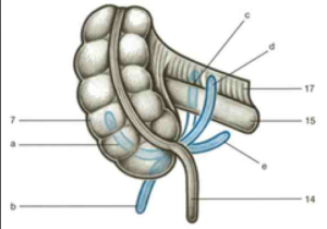 call them viral syndrome and discharge home? NO! So far, studies suggest that as opposed to the specificity, the sensitivity of this test is insufficient to RULE OUT the disease. Likely, the variation in the anatomical location of the appendix makes it difficult for us to always find it. While it usually arises 1cm inferior to the terminally ileum and moves inferior and medially, there is a great variation of ways it comes off of the terminal illeum.
So remember, if you see it, they probably have it, and if you don’t, well, continue monitoring with serial exams, and consider a surgical consult and CT scan if available.
And for even more information, check out the following resources:
Ultrasound Podcast – Appendicitis – multiple 20+ minute awesome podcasts
