Left LUNG
Right Lung
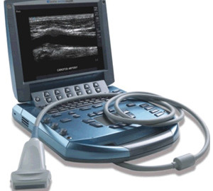
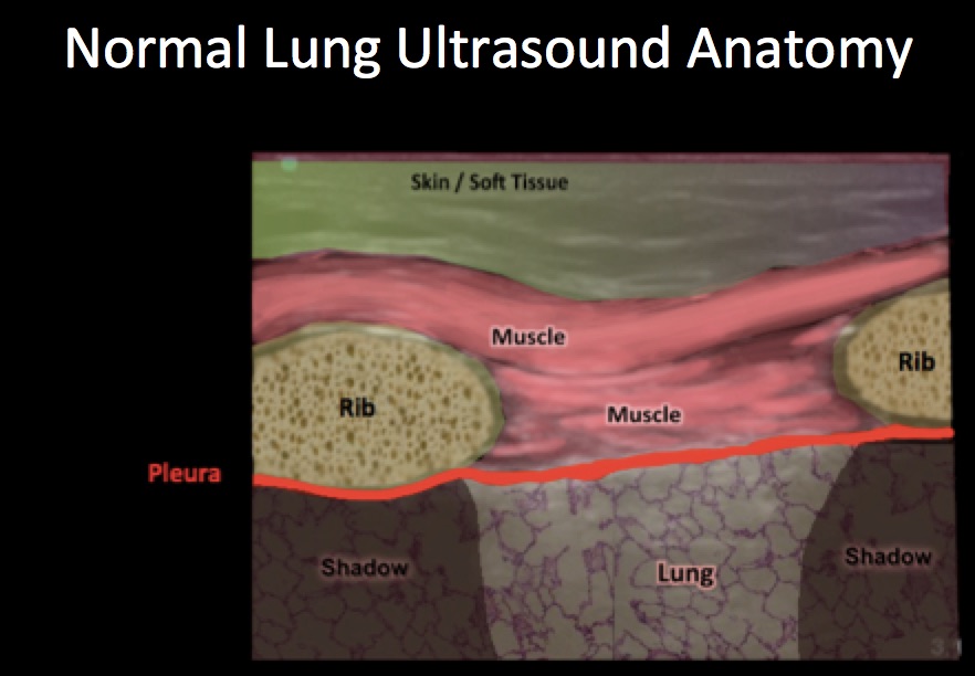
These ultrasounds performed by R1 Emergency Medicine resident Dr. DD at HUM, on a patient known to have TB and HIV who presented to the ED in Mirebalais complaining of lower left sided chest pain. The ultrasound of the left lung shows no “lung sliding.” Placing a linear probe vertically (marker toward the patient’s head) allows us to view the pleura between rib spaces. Like all bones, the ribs block ultrasound waves and therefore appear as dark shadows. Between these shadows is the pleura.
In a normal lung, you will be able to see the lung sliding back and forth against the pleura as the patient breaths, however this “lung sliding” will be absent if you are looking at a pneumothorax. Compare the video of the patient’s right lung (no pneumothorax, + lung sliding) and the left lung (with pneumothorax, – lung sliding) to see the difference.
When performing the ultrasound, Dr. DD correctly looked at the right lung first. Remember, whether it is an ear exam for otitis media or a lung exam with ultrasound, ALWAYS LOOK AT THE “GOOD” SIDE FIRST SO YOU KNOW WHAT TO COMPARE TO.
You can also use “M” mode, or “motion” mode, which provides an image showing tissue motion along a single ultrasound beam. When “m” mode is used on a normal lung, the lung (and the air inside it) moves back and forth across this single ultrasound beam creating a picture that is often compared to a sandy beach. In a pneumothorax, since there is no movement, using “m” mode will give the appearance of a “bar code.”

In the videos, Dr. DD uses m-mode nicely to demonstrate the presence of a pneumothorax.