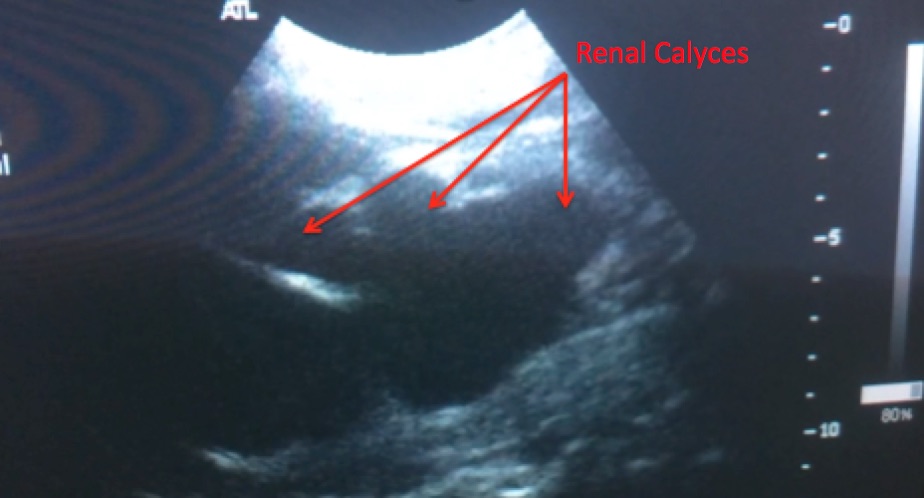
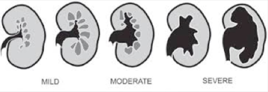
This ultrasound performed by R3 Family Medicine Resident Dr. DM of HSN demonstrates SEVERE hydronephrosis. This patient came through clinic and had minimal to no pain suggesting this was likely a chronic process. When estimating the degree of hydronephrosis, a general estimate based on the appearance of the renal pelvis is sufficient – there is no such thing as “40% hydronephrosis” or other exact numbers. A patient can have “mild” or “moderate” or “mild to moderate” or in the case of our patient, “severe” hydronephrosis.
This ultrasound also nicely demonstrates how important it is when evaluating for hydronephrosis to “fan” (keep the probe at the same spot and move it anterior and posterior) through the kidney. If you were to look at the kidney from just one view, the hydronephrosis could be mistaken for a large kidney cyst.
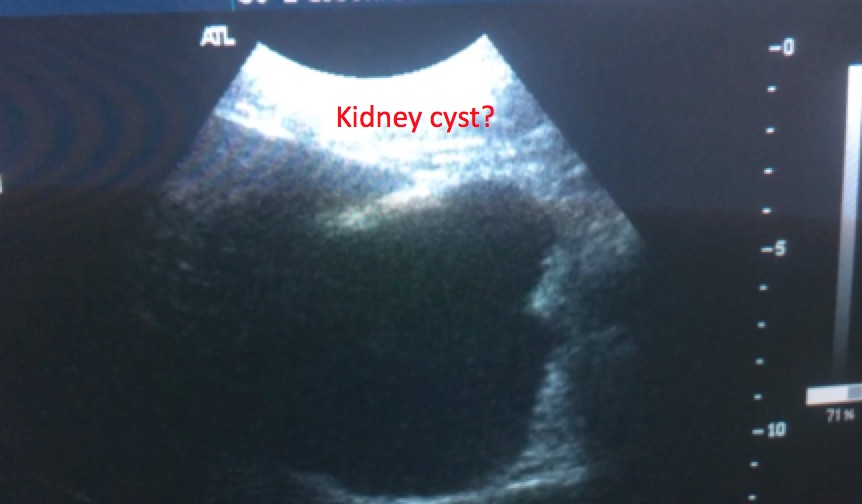
In future ultrasounds, you will see many kidney cysts, which are round “anechoic,” or black circles, usually at the edges of the kidney.
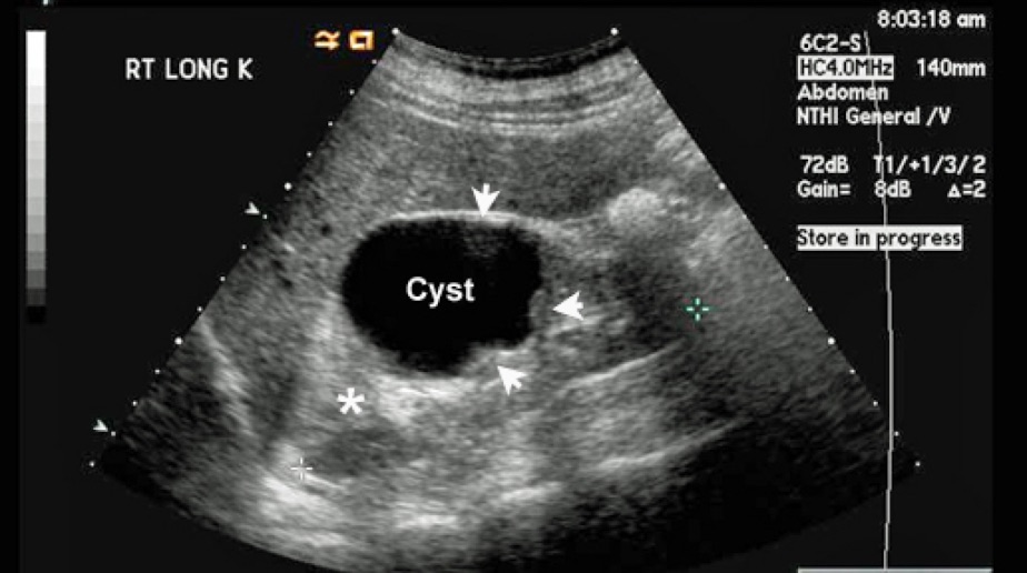
However when Dr. DM “fanned” through the kidney, he was able to clearly identify that the black “anechoic” structure was actually dilated renal calyces and therefore he correctly identified hydronephrosis.