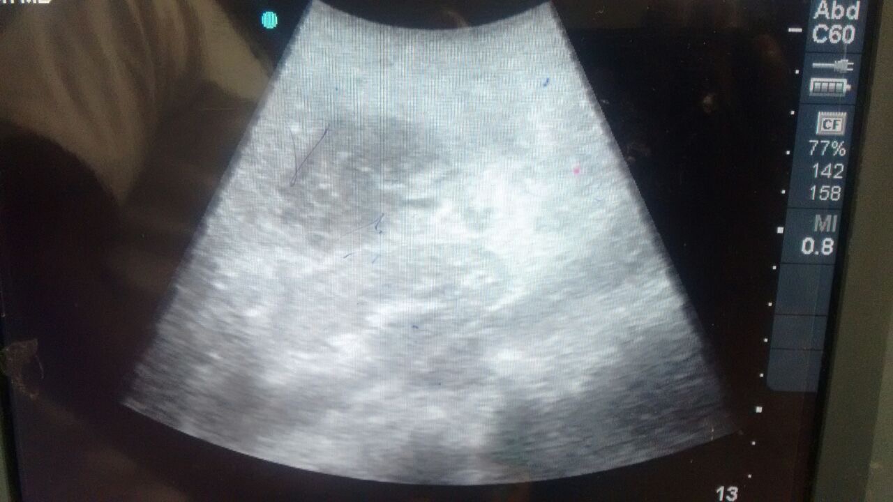
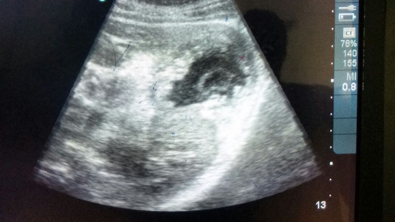
This ultrasound was performed by Dr. Menager, PGY-1 EM resident at HUM, on a 9 month old male with 9 days of abdominal pain and blood diarrhea. The study nicely demonstrates the “target sign,” the classic sonographic finding for intussusception.
Of note, this bedside study was performed in the PEDIATRICS ward. The patient did receive an ultrasound in the ED which did not show intussusception. At that time, the patient had intermittent pain. He was placed on ceftriaxone for presumed bacterial enterocolitis however his pain and bloody diarrhea continued and he was eventually admitted. On the ward, the patient continued to have pain and Dr. Menager, who was on his pediatrics rotation, cleverly decided to repeat the ultrasound study leading to the correct diagnosis. The patient was then taken to the operating room where the diagnosis of intussusception was confirmed and the appropriate treatment provided. GREAT WORK!
CLICK HERE TO SEE PAST VIDEO/IMAGES OF THE WEEK