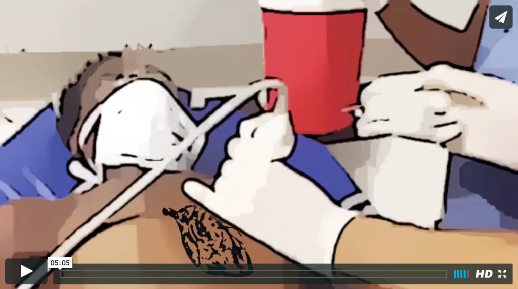
With high rates of tuberculosis, we really do see many pericardial effusions that require drainage. In fact, Dr. Plantin performed his second pericardiocentesis on his very next shift! Learn how to perform this procedure now so when the time comes for you to perform it, you will be ready
Category Archives: Ultrasound
How to place an IV line with ultrasound
Our ER nurses at HUM are awesome at obtaining IV access, but when even the best nurses cannot get that peripheral line, then being able to put in an IV line that will not infiltrate is an extremely useful skill for every Emergency Physician to have. What is the RIGHT WAY to perform this important ED procedure? Watch this video on EMin5 and find out!
Ultrasound – 10 – Intussusception
These ultrasounds, performed by EM residents at HUM, nicely demonstrate the classic sonographic finding consistent with intussusception: the target sign, or layers of invaginated bowel seen in cross section.
Ultrasound – 9 – Small bowel obstruction
These ultrasounds, performed by Dr. Ricot, R1 EM resident at HUM, nicely demonstrate sonographic findings consistent with a small bowel obstruction.
Ultrasound – 8 – Skull fracture
R2 EM resident Dr. Duneant, accompanied by one of our visiting professors, Dr. Aimee Tang, International Emergency Health Fellow at North Shore-LIJ, performed this ultrasound on a 2 yr old boy who was referred to the HUM ED from an outside hospital.
Ultrasound – 7 – Acute Appendicitis
A 25 yr-old female presented to HUM with RLQ pain and nausea. Megan Mickley, one of our awesome visiting professors, Ultrasound Fellow from Children’s Hospital of Philadelphia, performed this ultrasound with one of our residents, which nicely demonstrates acute appendicitis.